Nov. 25, 2015
 ASRT Foundation donors are supporting the future of the profession and providing a necessary avenue for research funding.
In May 2014 researchers Timmerie F. Cohen, Ph.D., R.T.(R)(T), CMD; Jeffrey S. Legg, Ph.D., R.T.(R)(CT)(QM); and Melanie C. Dempsey, M.S., R.T.(R)(T), CMD, FAAMD, received a research grant to study the effect of vertical off-centering during CT simulations on breast tissue exposure in Accelerated Partial Breast Irradiation patients. Donor funding is what makes grants like this available to researchers so that they can complete their investigations and move the profession forward.
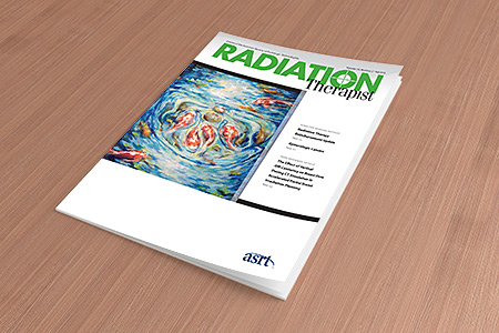
Thanks to their research grant, Timmerie, Jeffrey and Melanie completed their project and published their findings in the fall 2015 issue of Radiation Therapist. The following is an excerpt of that article.
“This study explored the effect of vertical off-centering during CT simulation on breast tissue exposure in APBI patients. When the table was lowered 3 cm, socenter anterior to midplane, breast exposure increased as much as 23%. A decrease of as much as 17% in exposure occurred when the table was raised 3 cm. These findings followed the same trend as previous research by Kaasalainen and colleagues.
Keeping in mind that breast tissue has a high sensitivity, radiologic technologists should avoid performing simulations on patients using a low table height to prevent increased dose to breast tissue due to misoperation of the automatic tube current modulation of the CT scanner. However, because the breasts are located anteriorly, increasing the table height also should be avoided to ensure breasts are included in the scan even though increasing table height decreases breast dose.
Every radiation science professional, including radiation therapists performing simulation, should consider radiation protection for their patients. Imaging that uses ionizing radiation is generally in the patient’s best interest, with the benefits outweighing the risks. This also is the case with APBI for the treatment of early, highly curable breast cancer. Regardless of the benefits, the stochastic effects of ionizing radiation should be acknowledged and reduced wherever possible. Even decisions that seem mundane, such as table height in a CT simulation, can affect sensitive tissues such as the breast.
Based on the results of this research, the authors recommend that radiation oncology departments ensure their CT simulators are used with proper optimization. Image optimization for APBI patients includes limiting scan range to the area of interest, avoiding vertical off-centering, and using the appropriate tube current modulation. Setting the isocenter at midplane (approximately the midaxillary line) should be the standard of care in CT simulation to avoid increased radiation to the breast.
Consideration also should be given to the subsequent use of ultrasonography to verify catheter placement in patients undergoing APBI after the initial CT scan used to verify catheter placement and to plan treatment. The Image Wisely campaign advocates “lowering the amount of radiation used in medically necessary imaging studies and eliminating unnecessary procedures.” Radiation dose from multiple CT scans in APBI treatment could be reduced by using ultrasonography in place of CT scans when verifying balloon placement daily.
This study has limitations. Only one type of CT simulator and one type of phantom were used. Measurements using multiple CT scanners across a variety of institutions and the use of multiple breast size phantoms would further support this research. A standard phantom cannot provide complete information regarding breast exposure because of differences in body and breast sizes. The measurement tool itself has limitations because the TLDs used have an overall uncertainty of 3.6% in their readings according to Hammer (oral communication, March 2015). Nevertheless, this study provides evidence that simulation techniques can affect radiation dose to sensitive tissues.”
As a Foundation donor, you are helping ensure that research is being conducted in the field by R.T.s, for R.T.s. You are creating a brighter future for the profession and improving patient care.